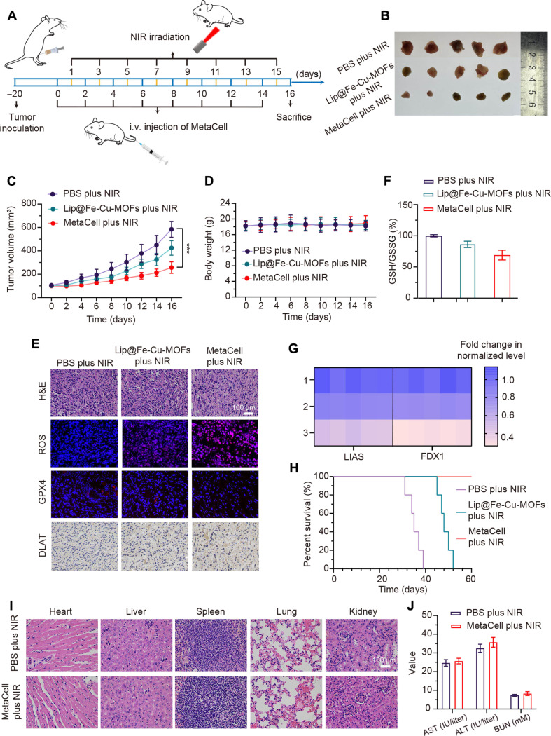
Fig. 6. Evaluation of in vivo antitumor capabilities of MetaCell.
(A) Therapeutic protocol illustrating MetaCell administration. (B) Visualization of tumor specimens following treatment. Mice were treated intravenously with PBS, Lip@Fe-Cu-MOFs [Fe (5 mg/kg)], or MetaCell (2 × 10⁷ cells per mouse) every 2 days, with a total of eight rounds of administration, followed by NIR stimulation (1.5 W/cm2, 808 nm) for 5 min, administered 24 hours postinjection. After the 16-day treatment, mice were euthanized, and tumors were subsequently harvested and imaged. (C) Tumor growth trajectories in 4T1 tumor–bearing mice post-MetaCell treatment. Tumor progression was monitored over a 16-day period. (D) Body weight fluctuations of 4T1 tumor–bearing mice over the 16-day treatment span. (E) Representative images of tumor sections stained with H&E, ROS, GPX4, and DLAT posttreatment. Mice were euthanized following the 16-day treatment period, and tumors were harvested, sectioned, and stained with H&E, ROS, GPX4, and DLAT. Scale bar, 100 μm. (F) Assessment of GSH depletion in tumor tissues: After a 16-day treatment regimen, euthanized mice’s tumors were excised, homogenized, and subjected to centrifugation. The GSH levels in the resultant supernatant were measured using a GSH and GSSG assay kit. (G) Capability of MetaCell to suppress FDX1 and LIAS expression. After 16-day treatment, 4T1 tumor–bearing mice from each group were euthanized, and tumors were harvested, homogenized, and centrifuged for subsequent LIAS and FDX1 expression assessment via quantitative real-time PCR (qRT-PCR). (H) Survival tracking of tumor-bearing mice over the course of the 60-day treatment in each group. (I) Histopathological evaluation via H&E staining of critical organ tissues after 16-day MetaCell plus NIR treatment. Scale bar, 100 μm. (J) Concentrations of ALT, AST, and BUN in mouse serum after 16-day MetaCell plus NIR treatment period. Data are represented as mean ± SD, (n = 5). ***P < 0.001 [one-way analysis of variance (ANOVA)].

