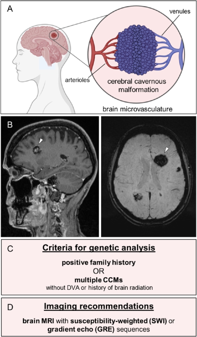
Figure 1.

Clinical features of CCMs. (A) CCMs are mulberry-like vascular lesions in the microvascular bed of the central nervous system. (B) Sagittal T1-weighted (left) and axial susceptibility-weighted magnetic resonance images (right) show a large CCM (white arrowhead) and multiple smaller CCMs in both hemispheres of a patient with a pathogenic CCM1 germline variant. (C) Criteria for genetic testing of patients with CCMs (adapted from [2]). DVA = developmental venous anomaly. (D) Recommended imaging techniques for diagnosis or follow-up of CCMs (adapted from [2]).
