Figure 3.
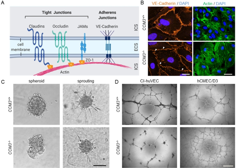
Disorganized cell junctions and impaired function in 3D models of angiogenesis upon CCM3 gene inactivation. (A) Scheme of endothelial tight and adherens junctions. ICS = intracellular space. ECS = extracellular space. ZO-1 = zonula occludens protein 1. JAM = junctional adhesion molecule. (B) In contrast to wild-type CI-huVECs, CI-huVECs demonstrated numerous small gaps (white arrowheads) and a less homogeneous pattern in VE-cadherin staining (red). Scale bar 20 µm. They also displayed significant actin stress fiber formation (green, phalloidin staining). Scale bar 25 µm. DAPI (blue) was used to stain cell nuclei. (C) CI-huVECs demonstrated impaired spheroid formation and VEGF-induced sprouting. The number and length of sprouts formed by spheroids upon stimulation with 25 ng/ml VEGF-A were significantly reduced. Scale bar 100 µm. (D) CCM3 gene inactivation had cell type-specific effects on endothelial tube formation on Matrigel. While tubes formed by CI-huVECs were unstable and had fallen apart 17 h after seeding on Matrigel, hCMEC/D3 cells formed more stable meshes than hCMEC/D3 wild-type controls. Scale bar 500 µm.

