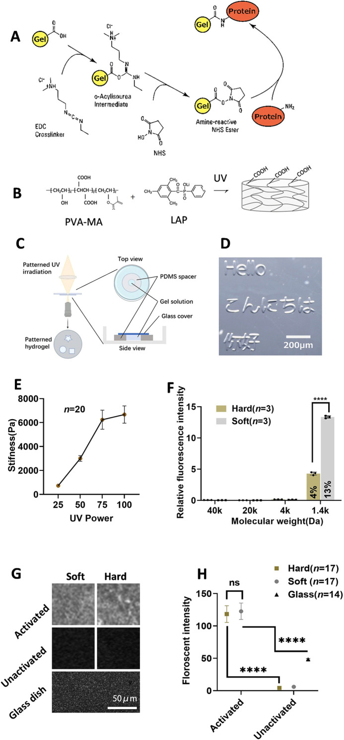
Fig. 1.

Photocurable PVA-PEG hydrogel. (A) The hydrogel can be activated to bond with proteins using 1-ethyl-3-(3-dimethylaminopropyl) carbodiimide/N-hydroxysuccinimide (NHS/EDC). (B) PVA-MA forms COOH-containing hydrogel upon ultraviolet (UV) irradiation. (C) Schematic for generating patterned hydrogel. (D) Patterned gel was generated using patterned UV irradiation, as shown in C. (E) Stiffness measurements for hydrogel generated using different UV doses. Data are mean±s.d. (n=20). (F) Fluorescent diffusion experiment: hydrogels were incubated with fluorescent molecules. Data are mean fluorescent intensity inside the gel relative to incubation solution±s.d. Black dots represent each sample (n=3). (G) Fluorescent protein-binding experiments. Activated and non-activated soft and hard gels, together with a glass-bottom dish, were incubated with fluorescent protein to evaluate the protein-binding ability. (H) Quantification of fluorescent protein binding experiments. Fluorescence intensity was measured using fluorescence microscopy (n=17 for hard, n=17 for soft, n=14 for glass). Data were obtained from at least three biological replicates. Data are mean±s.d. with the median values indicated by black horizontal lines. Scale bars: 200 µm in D; 100 µm in G. ns, not significant. ****P<0.0001(unpaired two-tailed t-test).
