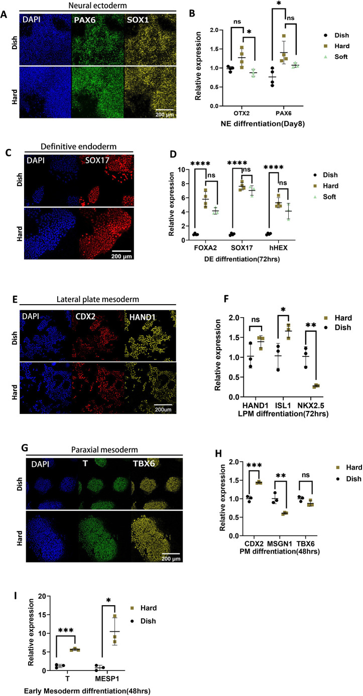
Fig. 4.
Differentiation of human pluripotent stem cells on gel. (A) Immunofluorescence of neural ectoderm differentiation. (B) qPCR of neural ectoderm differentiation. (C) Immunofluorescence of definitive endoderm. (D) qPCR of definitive endoderm differentiation. (E) Immunofluorescence of lateral plate mesoderm. (F) qPCR of lateral plate mesoderm differentiation. (G) Immunofluorescence of paraxial mesoderm. (H) qPCR of paraxial mesoderm differentiation. (H) qPCR of early mesoderm (BMP/bFGF) differentiation. Scale bars: 200 µm. Data were obtained from at least three biological replicates. Each dot represents an individual biological replicate, with black lines indicating median values. Data are mean±s.d. Statistical significance was determined using an unpaired two-tailed t-test. ns, not significant. ****P<0.0001, ***P<0.001, **P<0.01, *P<0.1.

