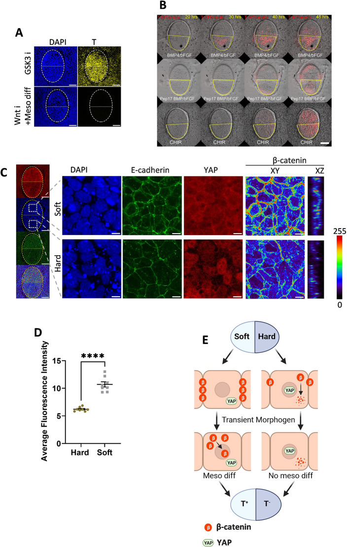
Fig. 6.
Speculated mechanisms of stem cell fate control. (A) Immunofluorescent staining of cells differentiated using GSK3 inhibition or by BMP/bFGF together with Wnt inhibition on a partially soft gel. The white dotted oval line indicates the whole gel pattern, with the upper half being soft and the lower half being hard. Scale bars: 100 µm. (B) Screenshot of live imaging of T-tdTomato reporter cells differentiated on a patterned gel using BMP/bFGF with CHIR (Movie 1) and using BMP/bFGF in the presence of Peptide 17 (Movie 2). The red color represents the tdTomato signal. The yellow line circles the soft part of the gel. Scale bar: 100 µm. (C) Immunofluorescent staining of hPSCs on a partially soft gel pattern before differentiation. The yellow dotted oval line indicates the whole gel pattern, with the upper half being soft and the lower half being hard. The white squares indicate the positions of the magnified views. The β-catenin channel is shown in XY and XZ section views as a heat map, with colors representing fluorescent intensity. Scale bars: 100 µm in the whole gel view; 10 µm in the magnified view. (D) Quantification of β-catenin fluorescent intensity based on z sections of immunofluorescent staining of the soft and hard regions on the same gel pattern. Each dot represents an individual measurement (n=8). Data were obtained from at least three biological replicates. Data are mean±s.d. ****P<0.0001 (unpaired two-tailed t-test). (E) Proposed mechanism of spatial cell fate control. On the soft gel, β-catenin formation is enhanced and YAP nuclear localization is inhibited, promoting sensitivity to Wnt signaling. On the hard region, cells have nuclear-localized YAP, which negatively impacts Wnt signaling. Together with downregulated β-catenin compared with the soft part, this leads to decreased mesoderm differentiation compared with the soft region.

