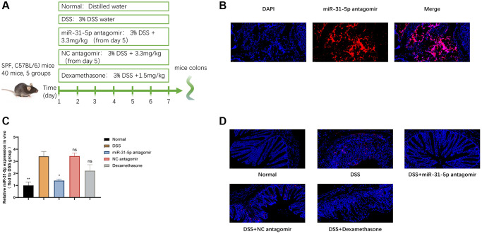
Figure 2.
miR-31-5p antagomir decreased the level of mir-31-5p in DSS-induced colitis. (A) Diagram of drug intervention process in animal experiments. (B) Immunofluorescence of frozen sections showed the localization of miR-31-5p antagomir in mouse colon. (C) QRT-PCR was used to analyze the level of miR-31-5p in colon tissues of mice. Each column represents the mean ± SD, n ≥ 3 from each group. ns represented P > 0.05, *P < 0.05, **P < 0.01 vs. DSS group. (D) In situ hybridization analysis of miR-31-5p in mouse colon. Red represented positive signal for miR-31-5p (magnification ×400).

