Fig. 3.
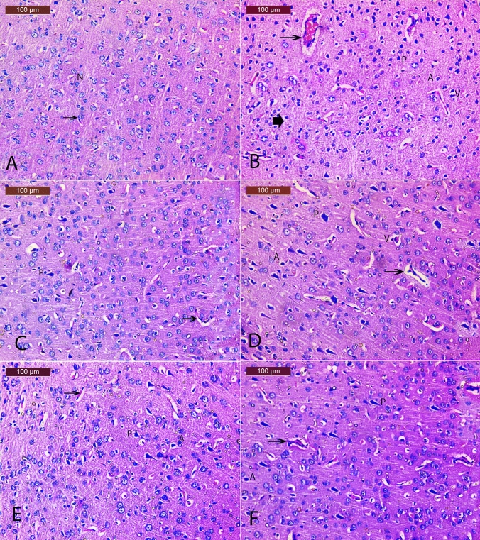
Effect of E. bonariensis on OVX/D-Gal-induced cerebral cortex histopathological changes. Representative H&E photomicrographs (cerebral cortex region) of all experimental groups (n = 2); magnification: Hand E × 200 A SO group showing normal structure of cerebral cortex and the neurons with their characteristic large vesicular nuclei (N); B OVX/D-Gal group showing numerous histopathological changes including a large number of damaged neurons, degenerated, necrotic (arrowhead), perineuronal vacuolation (V), pyknotic nuclei (P), apoptotic (A), and congestion of cerebral blood vessel (arrow); C Donepezil group showing nearly normal neuronal morphology with minimal pyknotic, apoptotic nuclei (P); D E. bonariensis 50 mg/kg group showing less histopathological changes except for pyknosis of some neurons (P), apoptotic nuclei, and perineuronal vacuolation (V); E E. bonariensis 100 mg/kg group showing almost normal histological structure. Some neurons had normally stained nuclei and other neurons showed minimal apoptotic cells (A) and pyknotic nuclei (P) with normal blood vessels (Bv); F E. bonariensis 200 mg/kg group showing almost normal neuronal cells of cortex with few histopathological changes such as minimal pyknotic (P) and apoptotic nuclei (A). Rats underwent either SO or OVX, and after 5 days, they received D-Gal (150 mg/kg/day, i.p) for 42 days. OVX/D-Gal-subjected rats were orally treated with donepezil (5 mg/kg/day) or the alcoholic extract of E. bonariensis at three different doses (50, 100, and 200 mg/kg/day) for 42 days, given 1 h prior to D-Gal. One day after behavioral testing (day 43), rats were decapitated, and the brains were separated for histopathological examination. OVX ovariectomy, D-Gal D-galactose, SO sham operation

