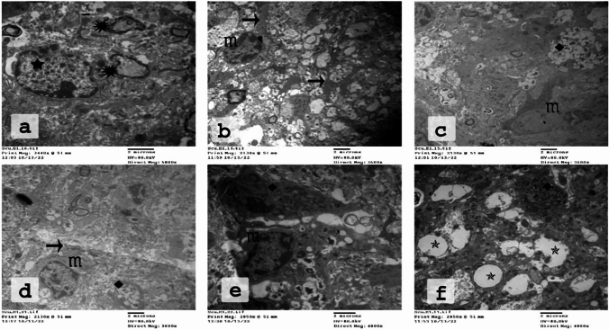
Fig. 5.
Electron micrographs of the CC of the CPZ-fed group showing stages of phagocytosis of myelin debris by microglia. Many distracted OLs were seen close to unhealthy nerve fibers. a Injured myelin (
 ) and destructed OLs (
 ) appear close to this injured myelin. b Microglia spread their processes ( →) among these axons. c–e Phagosomes with many debris (
 ). f Therefore, excessive demyelination and the accompanying phagocytosis might lead to numerous gaps and empty areas just next to microglia (
 ). (
n = 3 per group). CPZ; cuprizone, CC corpus callosum; OLs: oligodendrocytes

