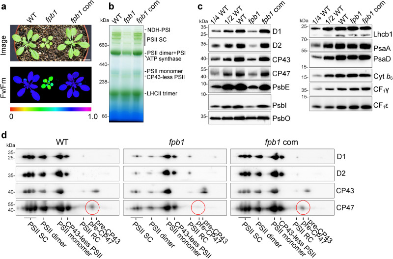
Fig. 1. Reduced level of PSII in fpb1.
a The image was taken from four-week-old plants grown in the greenhouse under about 50 μmol photons m−2 s−1 (top panel). Images of maximum quantum yield of PSII (Fv/Fm) were captured using a PAM imaging system. A false colour scale for Fv/Fm is shown at the bottom of the panel. fpb1 com, fpb1 mutant complemented with the FPB1 genomic sequence. Bar = 1 cm. b Blue native (BN)-PAGE analyses of the thylakoid protein complexes from four-week-old wild type (WT), fpb1, and fpb1 complemented (fpb1 com) plants. c Immunodetection of representative subunits of thylakoid protein complexes. Equal amounts of thylakoid proteins were separated by Tricine-SDS-PAGE (for PsbE, PsbI, and Cyt b6) or SDS-urea-PAGE (for others) and then probed with antibodies as indicated. A series of dilutions from WT was used for rough estimation of proteins. d 2D BN/SDS-urea-PAGE analyses of various PSII complexes from the fpb1 mutant. The pre-CP47 complex detected by CP47 antibody is surrounded by red circles. PSII SC, PSII supercomplexes. PSII RC, PSII reaction centre. Data are representative of two independent biological replicates. Source data are provided as a Source Data file.

