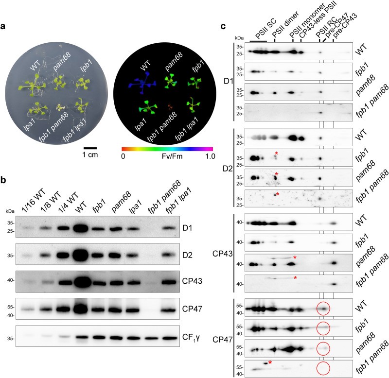
Fig. 3. Characterization of the fpb1 pam68 double mutant.
a Phenotypes (left) and Fv/Fm value (right) of 3-week-old fpb1, pam68, lpa1, fpb1 pam68, fpb1 lpa1 and WT plants grown on MS medium containing 3% sucrose under low light (20–30 μmol photons m−2 s−1). Fv/Fm values are indicated on a false colour scale. b Accumulation of PSII core subunits in three-week-old mutant and WT plants. Equal amounts of thylakoid proteins were loaded together with a series of dilutions of WT samples as indicated and separated by SDS-urea-PAGE. Immunoblotting with CF1γ antibody was used as a loading control. c Formation of the PSII assembly complexes in three-week-old fpb1, pam68, fpb1 pam68, and WT plants. To alleviate photodamage of the fpb1 pam68 double mutant, all the genotypes were grown under very low light irradiance (~5 μmol photons m−2 s−1). Thylakoid proteins were separated by 2D BN/SDS-urea-PAGE and probed with antibodies against D1, D2, CP43, and CP47. Non-specific signals are indicated by red asterisks. The pre-CP47 complexes detected with CP47 antibody are circled in red. PSII SC, PSII supercomplexes. PSII RC, PSII reaction centre. Data are representative of two independent biological replicates. Source data are provided as a Source Data file.

