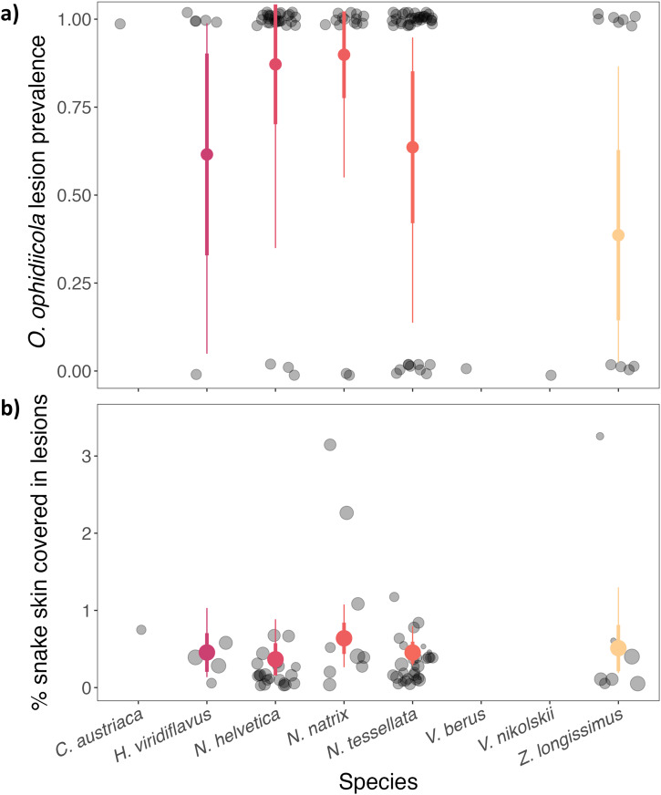
Fig. 3. Lesion prevalence and disease severity in O. ophidiicola-positive snakes across different species.
Color circles and whiskers show the model predicted posterior mean, ±standard deviation (thick lines), and 95% credible intervals (thin lines) for different species across all countries. a Each transparent black circle represents a single snake as being either negative (0) or positive (1) for presence of lesions, which was used to calculate the proportion of the population that tested positive (prevalence). b Each transparent black circle represents the percentage of the body of a single snake covered in lesions and the size of the circle is proportional to the total surface area of the snake (scale ranges 250–1000 cm2).

