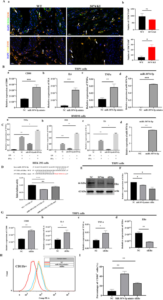
Fig. 2. MiR-3074-5p induced M1-polarization of macrophages by targeting ERα.
A (a) The representative images of multiplex immunohistochemistry staining of uterine tissue collected from a wild type pregnant mouse (WT) and a miR-3074-5p-knockin pregnant mouse (3074KI) at GD13.5; M1-subtype (F4/80+/CD86+) and M2-subtype (F4/80+/CD206+) macrophages (Mϕs) were stained in the paraffin sections, Orange: F4/80+, green: CD206+, yellow: CD86+; Scale bar = 20 μm; (b) the positive cell number of M1-subtype (F4/80+/CD86+) and M2-subtype (F4/80+/CD206+) Mϕs in 3074KI pregnant mice (n = 3) and WT pregnant mice (n = 3) at GD13.5. B Expression levels of CD80 (a), IL-6 (b), TNF-α (c) and miR-3074-5p (d) in THP1-derived Mϕs after 24 h of treatment with PMA (200 nM). Cells were transfected with negative control fragment (NC) or miR-3074-5p mimics (miR-3074-5p mimics) for 24 h, and then, mRNA levels of human M1-Mϕ markers (CD80, IL-6 and TNF-α) and miR-3074-5p were determined by RT-qPCR assay (n = 3 × 3). C Expression levels of TNF-α (a), iNOS (b), IL-6 (c) and miR-3074-5p (d) in Bone marrow-derived macrophages (BMDMs). Cells were transfected with NC or miR-3074-5p mimics for 24 h, and then, treated with LPS and IFNγ for another 24 h, mRNA expression levels of mouse M1-Mϕ markers (TNF-α, iNOS and IL-6) and miR-3074-5p were determined by RT-qPCR assay (n = 3 × 3). D Upper: Schematic representation of ERα 3ʹUTR sequence demonstrating predicted putative and mutated miR-3074-5p binding site (mutations in red); below: Relative luciferase activity in HEK293 cells co-transfected by negative control plasmid and wild type of ERα-3ʹUTR sequence (NC+3ʹUTRwt), or co-transfected by the miR-3074-5p overexpression plasmid and wild type ERα-3ʹUTR sequence (miR-3074OE+3ʹUTRwt), or co-transfected by the miR-3074-5p overexpression plasmid and mutant 3ʹ-UTR sequence of ERα (miR-3074OE+3ʹUTRmut) respectively. E Representative images of Western blotting detection of ERα protein expression levels in THP1 cells. Cells were respectively transfected by NC fragment (NC), miR-3074-5p mimics (3074 m), or ERα specific-siRNA (siERα). F Relative expression level of ERα protein in THP1 cells. Cells were respectively transfected by NC fragment (NC), miR-3074-5p mimics, or ERα specific-siRNA (siERα) for 48 h, and then, ERα expression level was detected by Western blot assay (n = 3). G Expression levels of CD80 (a), IL-6 (b) and TNF-α(c) and ERα (d) in THP1 cells. Cells were respectively transfected with negative control fragment (NC) or ERα-siRNA (siERα) for 24 h, and then, mRNA levels of M1 polarization markers (CD80, IL-6 and TNF-α) and ERα were determined by RT-qPCR (n = 3 × 3). H Flow cytometry assay of CD11b-FITC and CD80-PE on cell surface of THP1 cell. I The percentage of CD80-positive cells in the CD11b-positive cell population of THP1 cells transfected respectively by NC, miR-3074-5p mimics or siERα (n = 3 × 3). All data are shown as the mean ± SEM, *p < 0.05, **p < 0.01, ***p < 0.001, ns: p > 0.05.

