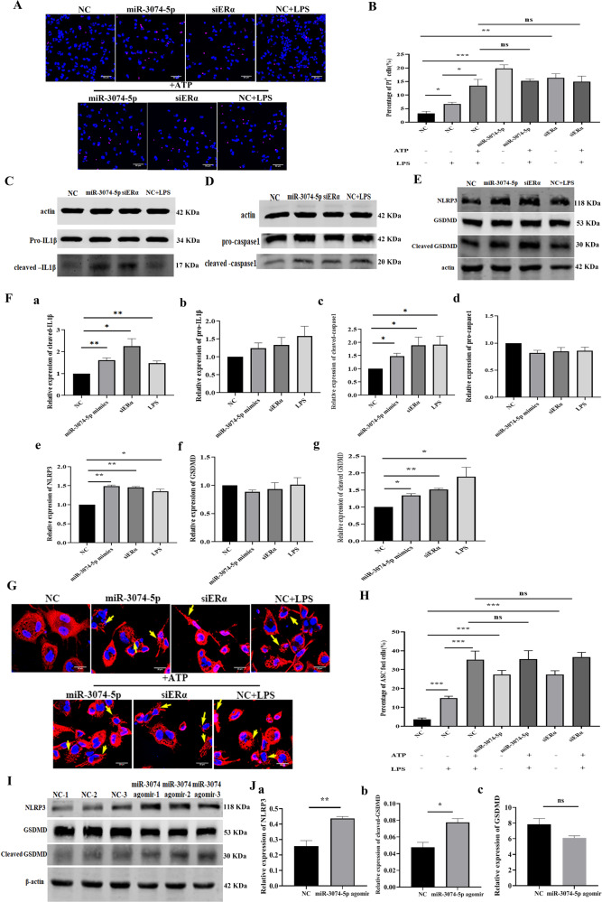
Fig. 3. MiR-3074-5p promoted pyroptosis of THP1-derived Mϕs and uterine expression levels of pyroptosis-related proteins.
A Representative images of propidium iodide (PI) staining.THP1-derived Mϕs were respectively transfected with negative control fragment (NC), miR-3074-5p mimics (miR-3074-5p) or ERα-siRNA (siERα), or treated with LPS (200 ng/ml) for 4 h and then treated with 5 mM ATP for 30 min to induce pyroptosis (red: PI, blue: hoechst33342, scale bar = 50 μm). B The percentage of PI-staining positive cells in total cells. PI-positive cells were counted on THP1-derived Mϕs from at least three independent fields per group. C Representative images of Western bolt assay of pro- and cleaved-IL1β proteins. D Representative images of Western bolt assay of pro- and cleaved-caspase1 proteins. E Representative images of Western bolt assay of NLRP3, GSDMD, and cleaved-GSDMD proteins. F The relative expression levels of cleaved-IL1β (a), pro-IL1β (b), cleaved-caspase1 (c), pro-caspase1 (d), NLRP3 (e), GSDMD (f), and cleaved-GSDMD (g) in THP1-derived Mϕs detected by Western blot assay. Cells were respectively transfected with NC fragment (NC), miR-3074-5p mimics (miR-3074-5p) or ERα-siRNA (siERα), or treated with LPS (200 ng/ml) (n = 3). G Representative images of immuno-fluorescence (IF) staining of ASC in THP1-derived Mϕs (red: Cy3-conjugated secondary antibody against ASC antibody, blue: hoechst33342, scale bar = 20 μm). H The percentage of THP1-derived Mϕs forming ASC aggregates. Cells were respectively transfected with negative control fragment (NC), miR-3074-5p mimics (miR-3074-5p) or ERα-siRNA (siERα), or treated with LPS (200 ng/ml) for 4 h and then treated with 5 mM ATP for 30 min to induce pyroptosis (n = 3 × 3). I Representative images of Western blot assay of uterine expression levels of NLRP3, GSDMD and cleaved-GSDMD protiens in GD8.5 pregnant mice. J Relative uterine expression levels of NLRP3 (a), GSDMD (b) and cleaved-GSDMD (c) protiens in mice detected by Western blot assy. Pregnant mice were respectively treated with NC fragment (n = 3) or miR-3074-5p agomir (n = 3), and the uterine tissue was collected at GD8.5. All data were presented as mean ± SEM. *p < 0.05, **p < 0.01, ***p < 0.001, ns: p > 0.05.

