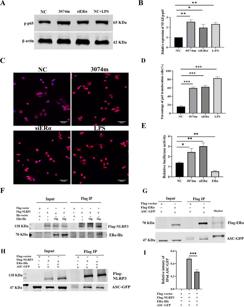
Fig. 4. MiR-3074-5p activated NLRP3 inflammasome by targeting ERα in macrophages.
A Rrepresentative images of Western blot assay on expression level of phosphorylated NFκB/p65(S276) protein in THP1-derived Mϕs. B Relative expression level of phosphorylatedNFκB/p65 (S276) protein in THP1-derived Mϕs detected by Western blot assay (n = 3). C Representative images of confocal immunofluorescence microscopy analysis of the NF-κB/p65 proteins translocated into the nuclei of THP1-derived Mϕs. D Percentage of cells with NFκB/p65 nuclei translocation (n ≈ 3 × 50 cells). E NFκB/p65 luciferase reporter activity relative to negative control in HEK293 cells following indicated treatments (n = 3). F HEK293T cells were co-transfected with different concentration of ERα-His plus Flag-NLRP3 or Flag vector for 24 h. Cell lysates were immunoprecipitated using anti-Flag beads and analyzed using anti-His and anti-Flag antibody. G HEK293T cells were co-transfected with ASC-GFP plus Flag-ERα or Flag vector for 24 h. Cell lysates were immunoprecipitated using anti-Flag beads and analyzed using anti-GFP and anti-Flag antibody. H HEK293T cells were co-transfected with ERα-His plus Flag-NLRP3 or Flag vectorand ASC-GFP for 24 h. Cell lysates were immunoprecipitated using anti-Flag beads and analyzed using anti-GFP and anti-Flag antibody. I The quantification data analysis of ASC-GFP immunoprecipitated by anti-Flag beads (n = 3). NC: cells were transfected with NC fragment; miR-3074-5p: cells were transfected with miR-3074-5p mimics; siERα: cells were transfected with ERα-siRNA; LPS: cells were treated by LPS (200 ng/ml); ERα: cells were transfected with ERα-expression recombinant plasmid. All data are shown as the mean ± SEM. *p < 0.05, **p < 0.01, ***p < 0.001.

