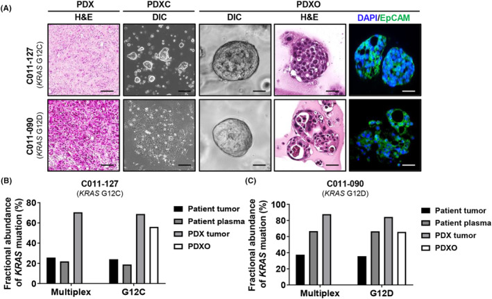
FIGURE 3.

Characterization of patient‐derived xenograft (PDX), PDX‐derived cell (PDXC), and PDX‐derived organoid (PDXO) from percutaneous liver biopsy tissue from a patient with metastatic pancreatic cancer. (A) Representative images of PDX tumor tissue and organoid using hematoxylin and eosin (H&E) staining and immunofluorescence. EpCAM‐positive cells are stained green. The nucleus was stained blue by DAPI staining. Scale bar = 50 μm. (B, C) Comparison of KRAS mutation fractional abundances obtained using droplet digital PCR (ddPCR) with specific primers for G12D or G12C mutation, among organoid and patient, PDX tumor tissues or plasma. A multiplex KRAS mutation detection kit for seven common KRAS mutations (G12A, G12C, G12D, G12R, G12S, G12V, and G13D) was used. Genomic DNA was purified from formalin‐fixed paraffin‐embedded (FFPE) tissue of F0 and F1. Purified DNA was used for ddPCR with KRAS screening.
