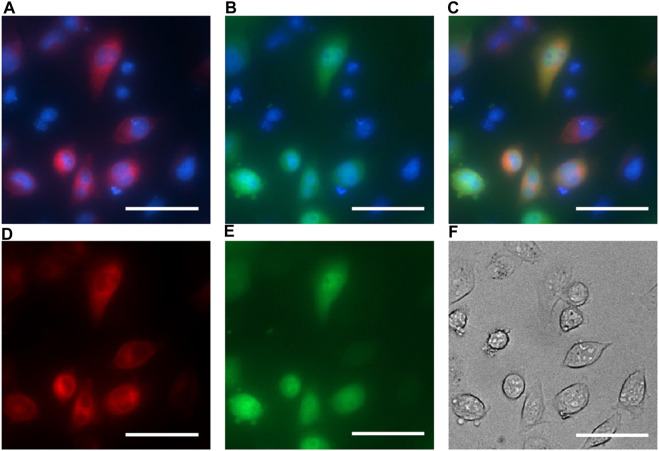
FIGURE 2.
EPG BRET fluorescent imaging for cell localization. HeLa cells co-transfected with the EPG BRET construct and EPG N terminus HaloTag construct and imaged with ×40 magnification. Hoechst dye was used as a nuclear marker and imaged using the DAPI filter (blue: A, B, and C). The EPG HaloTag construct was imaged using a JFX 650 dye with the Cy5 filter overlaid with a nuclear marker (A) and without a nuclear marker (D). EPG BRET construct was imaged using the GFP filter overlaid with a nuclear marker (B) and without a nuclear marker (E). Merged image of the three channels (C) shows an expression of the EPG BRET construct in the cytoplasm, and the EPG HaloTag construct on the cell membrane. (F) Phase contrast image of cells. Scale bar = 50 µm.

