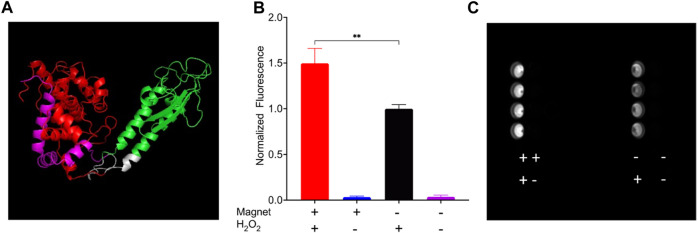
FIGURE 4.
HEK 293FT cells expressing EPG split APEX2 show an increase in fluorescence in response to magnetic field. All wells were treated with Amplex UltraRed reagent and the four combinations of with or without magnetic stimulus and H2O2 for 30 min. (A) Predicted structure of EPG split APEX2 with EPG (green), AP fragment (Red), EX fragment (magenta), and linkers (white). (B) Endpoint results of cells treated with all combinations of static magnetic stimulus and hydrogen peroxide (n = 4 independent experiments). (C) Image of a plate taken with a Cy3 filter after experiment for the detection of resorufin accumulation. Statistical analysis was performed using an unpaired t-test with Welch’s correction. The (**) denotes p-value <0.01.

