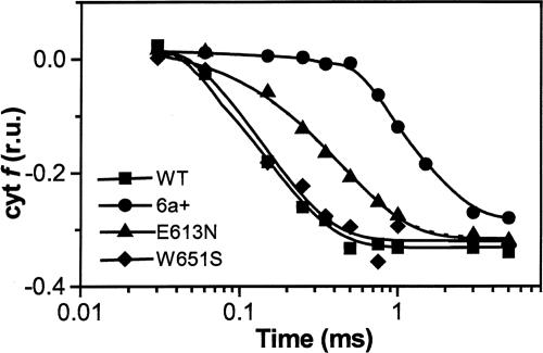
Fig. 2.
In vivo cyt f oxidation kinetics in WT and mutant Chlamydomonas cells. Cells were harvested during exponential growth (≈2 × 10⁶ cells per ml) and resuspended in minimum high salt medium (19), with addition of 20% (wt/vol) Ficoll to prevent cell sedimentation. Kinetics were obtained by deconvolution of absorption changes at 545, 554, and 573 nm after excitation with a laser flash, as described in Materials and Methods. Traces were normalized to the maximum amplitude of the cyt f oxidation signal; r.u., relative units. The duration of the actinic laser flash and of the detecting light-emitting diode flash was 10 ns and 10 μs, respectively.

