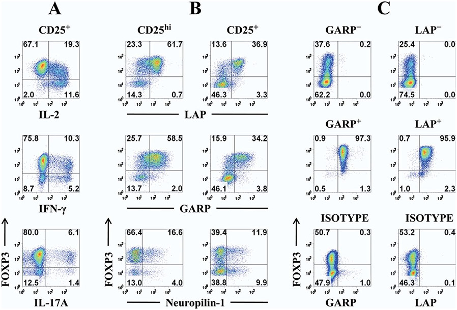
Fig. 1.
Selective expression of LAP and GARP on activated human Tregs in expansion cultures allows their repurification. (A) CD25+ cells were purified with magnetic beads and expanded in vitro by stimulation with anti-CD3/CD28 Dynabeads and 100 U/ml IL-2 for 14 days. The cells were then restimulated for 5 hours with phorbol myristate acetate/ionomycin in the presence of brefeldin A and analyzed for expression of FOXP3 (clone 236A/E7) and intracellular cytokines. (B) Human FACS-sorted CD4+CD127lowCD25hi (CD25hi) and CD25+ bead purified cells were expanded in vitro for 12 days, restimulated for 48 hours with anti-CD3/CD28, and then stained for surface LAP (clone 27232, R&D Systems), GARP (clone Plato-1, Alexis Biochemicals) or Neuropilin-1 (clone 446921, R&D Systems) expression and intracellular FOXP3. (C) CD25+ cells were expanded in vitro for 12 days and then restimulated for 48 hours with anti-CD3/CD28. Purified LAP+ and LAP− or GARP+ and GARP− fractions were isolated from the expanded cells using magnetic beads against LAP or GARP. Surface GARP, LAP, or isotypes and intracellular FOXP3 staining was performed.

