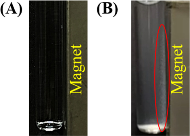
Figure 1.

Examination of the magnetic Eu3+–bacterium conjugates. Photographs of the (A) blank sample (0.39 mL) containing Tris buffer at pH 8 only and (B) the bacterial samples (0.39 mL, 0.5 mg mL–1) containing S. aureus prepared in Tris buffer (pH 8) obtained with the addition of Eu3+ (75 mM, 10 μL), followed by microwave heating (power: 180 W) for 2.25 min and magnetic isolation by placing an external neodymium magnet (∼4000 G). The photographs were taken under room light. The red oval indicates the location of the magnetic Eu3+–bacterium conjugates.
