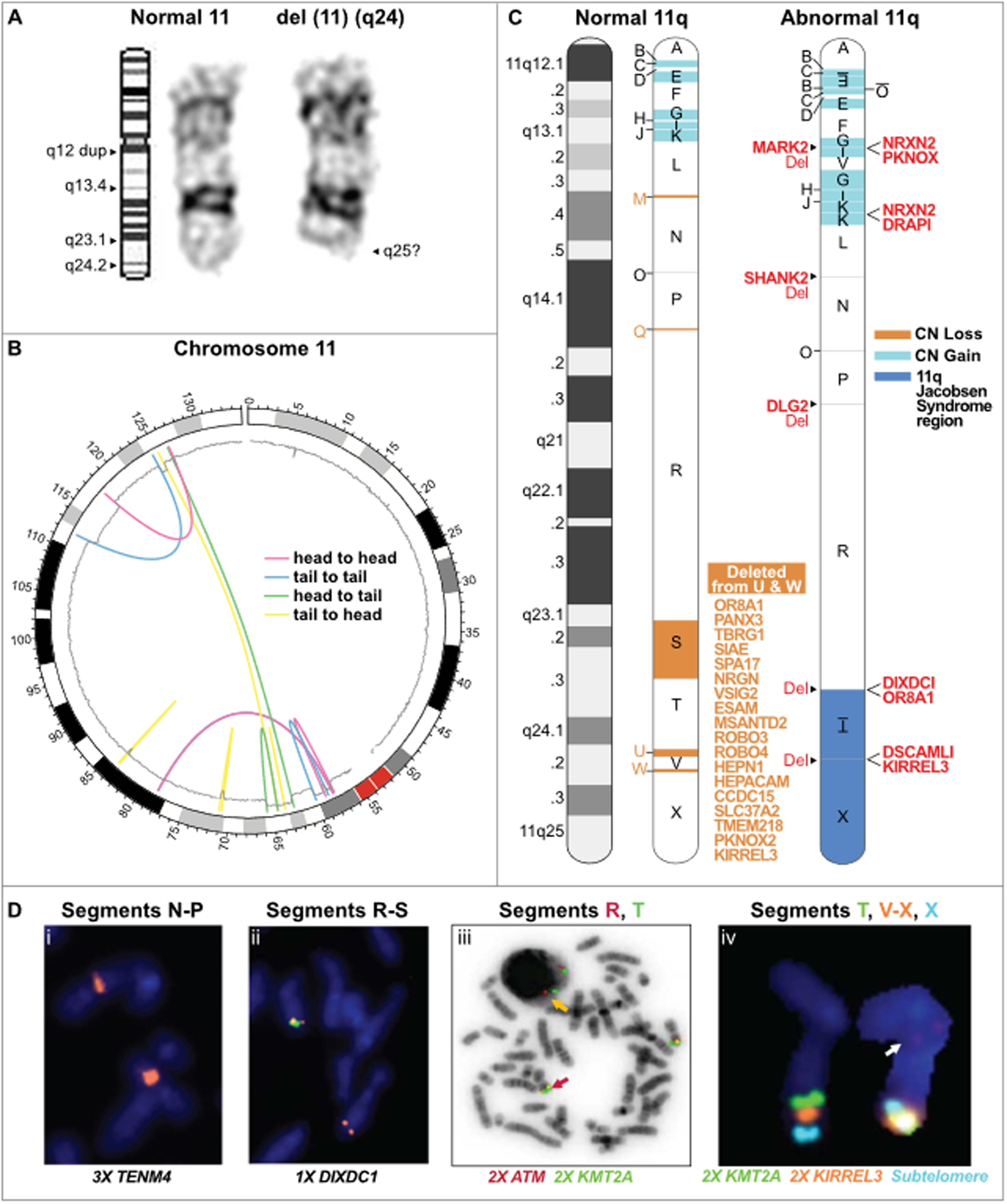
Figure 2.

Chromoanasynthesis of chromosome 11q as a mechanism for terminal 11q deletions in a patient with Jacobsen syndrome. (A) G band cytogenetic analysis of chromosome 11 in a Jacobsen Syndrome patient. Left, ideogram of normal chromosome 11 with labeled q arm bands including location of a q12 duplication (dup); middle, normal copy of patient chromosome 11; right, abnormal copy of patient chromosome 11 with deletion of band q24. Results are representative of 10 individual metaphases. See Figure S1 for the complete G-banded karyotype and spectral karyotyping. (B) Circos plot summarizing the rearrangements of the derivative chromosome 11. The chromosome is arranged circularly end-to-end. The outermost ring represents the chromosome ideogram with the centromere indicated in red. The inner ring represents the copy number based on smoothed copy number values from the chromosome microarray data. Centrifugal and centripetal deviations of the copy number tracing mark regions of gain and loss, respectively. The colored lines indicate the position and orientation of the breakpoint junctions based on the paired-end read data. The orientation of the rearrangements are head to head (red); tail to tail (blue); head to tail (green); or tail to head (yellow), as indicated in the inset.
(C) Linear model of the derivative chromosome 11q arm. Left, normal chromosome 11q depicted as a G-banded ideogram (left) and as a segmented chromosome (right) with letter-named segments (A-X) whose boundaries are the breakpoints defined by whole genome sequencing of patient DNA. Right, model of derivative chromosome 11q. Deleted segments are in salmon; duplicated segments are in blue; inverted segments are underlined and written upside down. Segment O possibly had 3 copies based on reads in the Integrative Genomics Viewer. Genes that are partially deleted (Del) or fused are listed in red adjacent to their position on the derivative chromosome. The 18 genes deleted from segments U and W in the Jacobsen region are listed in salmon.
(D) Fluorescence in-situ hybridization of patient metaphase spreads using specific probes for the genes indicated at the bottom of each panel located in the segments indicated at the top of each panel. The copy number state is indicated by #X, next to the corresponding gene. Panels iii and iv show results of combined FISH for the indicated probes which are color-aligned with the segment names. In panel iii, the orange arrow points to the wild type arrangement of KMT2A/MLL (green signal) and ATM (red signal). In panel iv, the wild type signal arrangement of chromosome 11q is on the left and the abnormal arrangement is on the right. Evidence of segment V translocation is visible (arrow).
