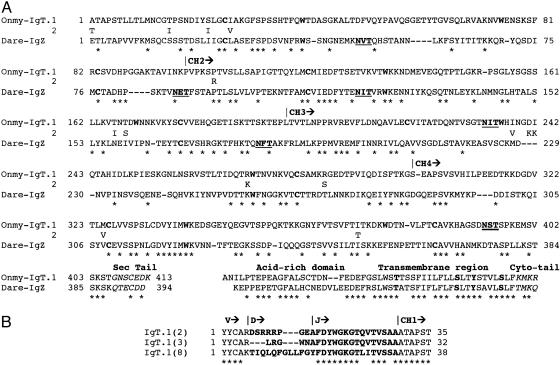
Fig. 1.
Comparative analysis of IgT in teleost fish. (A) Amino acid alignment of the rainbow trout (Onmy) IgT and zebrafish (Dare) IgZ C domains. C regions (CH) are based on cDNA and germ-line sequences for rainbow trout. Cysteine and tryptophan residues typical of the Ig fold are in boldface type. Potential N-linked glycosylation sites are underlined, and residues differing between the IgT duplicates are shown immediately under the IgT.1 sequence. * denotes identity, and – indicates gaps. S, T, and Y residues found in the transmembrane region typical of associating with B cell coreceptors CD79A/B are in boldface type. (B) CDR3 junctions for three IgT VDJ rearrangements.

