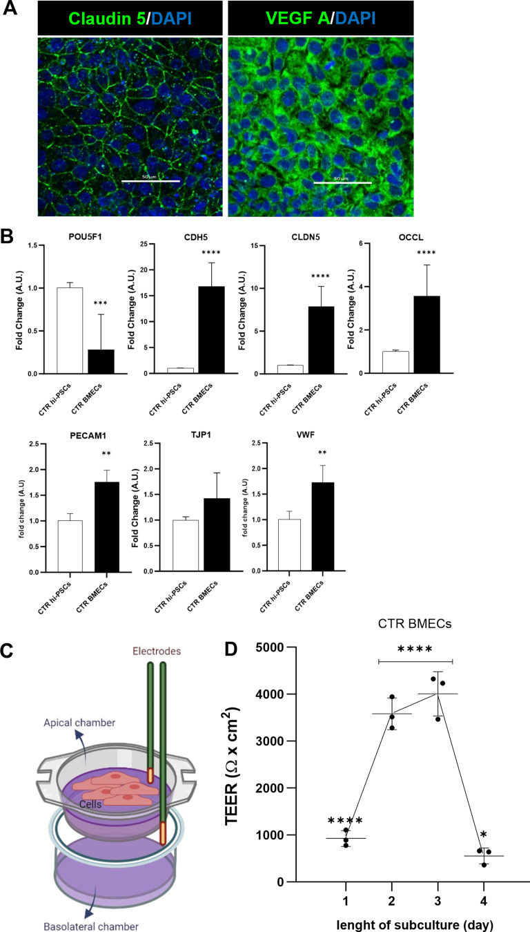
Fig. 1.
BMEC-like cells can be differentiated from hi-PSCs and can produce a cell barrier in vitro. A) Immunocytochemistry of BMEC cells differentiated from a healthy hi-PSCs donor. Claudin-5 and Vascular endothelial growth factor A (VEGF-A) using the RA-enhanced Lippmann’s laboratory protocol from 2019 [19]. Images were acquired with a Nikon confocal microscope. Scale bar 50 μm. B) Transcriptional expression of pluripotency and endothelial-specific markers in hi-PSCs and derived BMEC-like cells. Hi-PSC marker: POU5F1 (OCT4). Tight junctions: CDH5 (VE-CADHERIN), CLDN5, OCLN, PECAM1 (CD31), TJP1 (ZO-1) and VWF. The qRT-PCR data are plotted as mean ± s.d. Student’s unpaired t-test (****p < 0.0001), N = 3. C) Schematic representation of transendothelial resistance measurement (TEER). Cells plated on a permeable Transwell™ insert (0.4 μm PET) were assessed for TEER with an EVOM2 stx2 electrode. The diagram was created on Biorender.com D) BMEC passive barrier as shown by TEER following subculture for healthy donor hi-PSCs. Error bars represent the standard deviation of triplicate Transwell™ filters (N = 3). Statistical significance was determined using One-Way ANOVA (****p < 0.0001). N = 3

