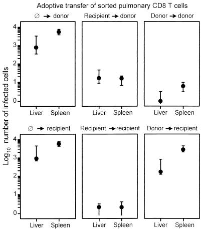
FIG. 10.
MHC genotype-specific, differential antiviral function of sorted donor-derived and recipient-derived pulmonary CD8 T cells. The results of the histological ISH analyses shown in Fig. 9 for selected 0.05-mm2 areas of tissue were substantiated by counting the numbers of infected cells in representative 10-mm2 areas of liver and spleen sections. Closed circles represent median values, and bars indicate ranges of the counted cell numbers for three adoptive transfer recipients per experimental group.

