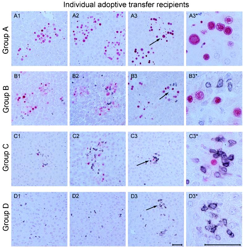
FIG. 3.
Two-color immunohistological analysis of liver tissue infection and T-cell infiltration. Pulmonary lymphocytes derived from lung infiltrates of infected chimeras at 5 weeks after BMT were tested for their antiviral and protective ability by intravenous adoptive transfer into infected BALB/c indicator recipients that were immunocompromised by 6 Gy of γ irradiation. The analysis was performed on day 14 after cell transfer. Group A, controls given no pulmonary T cells; groups B, C, and D, recipients of adoptive transfer of 104, 105, and 106 pulmonary lymphocytes, respectively. Panels A1 through D3 represent liver sections from individual recipients, three per experimental group. Infected cells, mostly hepatocytes, are identified by red nuclear staining of the IE1 protein pp89 of murine CMV. Infiltrating T cells are identified by black staining of CD3ɛ. Arrows mark areas of interest shown in overview (A3 through D3) and resolved to greater detail (A3* through D3*). Note the cytomegalic cells with prominent intranuclear inclusion bodies in panel A3* and the inflammatory foci visible under the conditions of group C. Photographs cover 0.08 mm2 (overview panels) and 0.005 mm2 (detail panels) of liver sections. The bar markers represent 50 μm.

