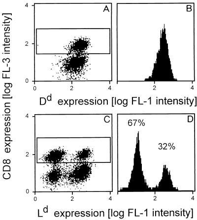
FIG. 6.
Chimerism among CD8 T cells in pulmonary infiltrates. Pulmonary lymphocytes were isolated at the peak of infiltration after BMT (6 Gy, 107 BMC) and infection. Three-color cytofluorometric analysis was performed for the combination FITC (FL-1)-Dd or FITC (FL-1)-Ld plus PE (FL-2)-TCR α/β plus RED613 (FL-3)-CD8. A gate was set on lymphocytes, and the analysis was restricted to α/β T cells by a second gate set on positive FL-2. (A and C) Two-dimensional dot plots of FL-3 (CD8) versus FL-1 (Dd [A] and Ld [C]) for 20,000 α/β T cells. CD8-positive cells are marked by a frame. (B and D) Histograms of FL-1 for the framed FL-2- and FL-3-positive CD8 T cells. The percentages of Ld-negative, recipient-derived and Ld-positive, donor-derived cells are indicated.

