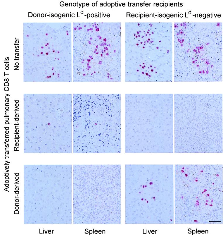
FIG. 9.
Control of viral replication in tissues of adoptive transfer recipients by sorted donor-derived and recipient-derived pulmonary CD8 T cells. The in vivo antiviral activity of the sorted CD8 T cells was tested by adoptive transfer of 50,000 cells into immunocompromised, γ-irradiated (6 Gy) indicator recipients, which were chosen to be isogenic either with the original BMT donor (BALB/c) or with the original BMT recipient (BALB/c-H-2dm2). Virus replication in liver and spleen of the indicator recipients was monitored on day 14 after the cell transfer by ISH specific for a gB gene DNA sequence of murine CMV. The red staining visualizes infected cells identified by the accumulation of viral DNA in an intranuclear inclusion body. Each photograph covers a 0.05-mm2 area of tissue section. The bar marker represents 50 μm.

