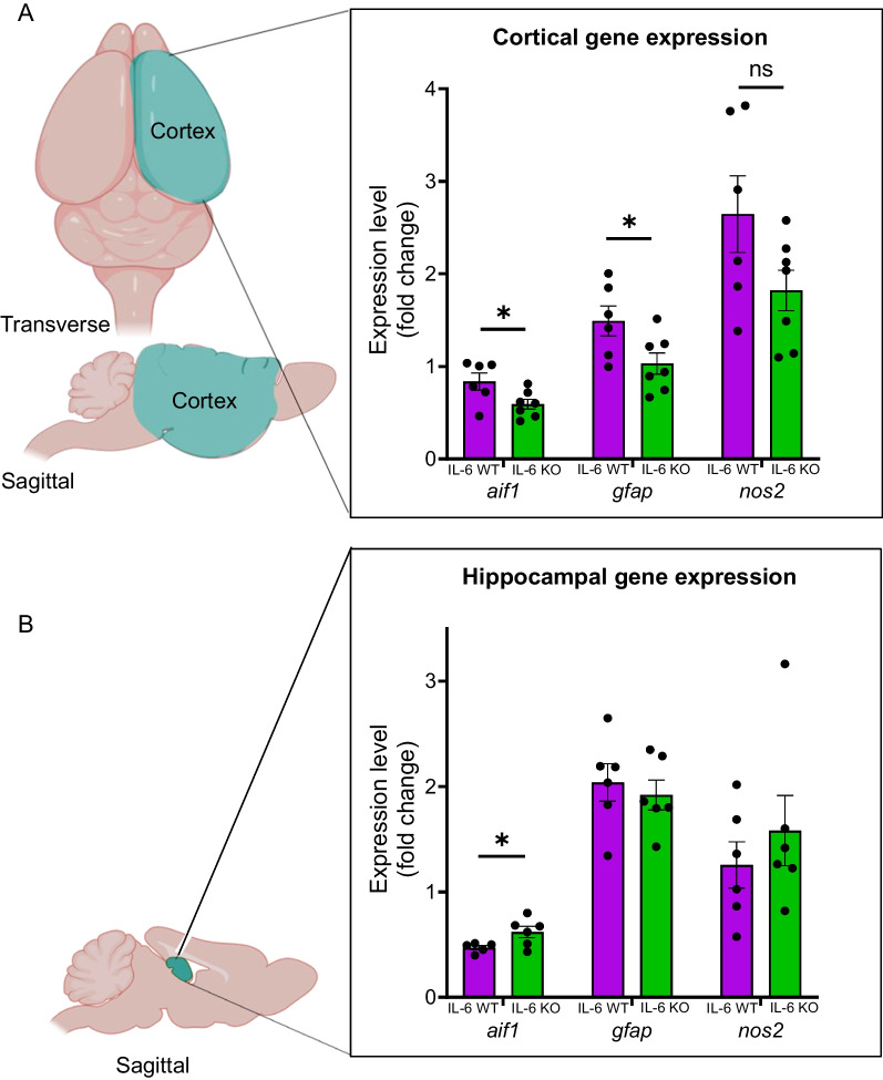
Fig. 5.
Cortical expression of glial genes is reduced in IL-6 KO MRL/lpr mice. Bulk gene expression was quantified in a whole-hemisphere of cortex (A; blue region on diagram) and the unilateral hippocampus (B; blue region on diagram) from IL-6 KO mice and compared to that of IL-6 WT mice. The reference sample was cortex, collected from a single sex and age-matched MRL/mpj mouse. One of the two separate cohorts (Cohort A) was allocated to quantification of brain gene expression using real time quantitative PCR, while the other (Cohort B) was reserved for subsequent histologic analysis. Measured genes included aif1 (allograft inhibitor factor 1; Iba-1 protein) which reflects monocytic and microglial activation/proliferation, gfap (glial fibrillary acidic protein) which reflects astrocytic activation/proliferation, and nos2 (nitric oxide synthase 2) which is a producer of inflammatory nitric oxide. *p < 0.05

