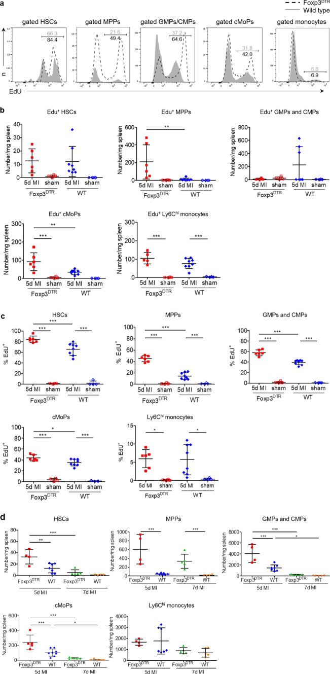
Fig. 3.
Proliferation analysis and quantification of precursor cells and monocytes in wild type (WT) and Foxp3DTR mice after myocardial infarction. a, b, c In vivo proliferation of HSCs, MPPs, GMPs/CMPs, cMoPs, and monocytes in the spleen 5 days after MI in wild type vs. Foxp3DTR mice. Representative plots a and quantitative data b, c for intracellular EdU analysis in MI animals. d Quantification of HSCs, MPPs, GMPs/CMPs, cMoPs, and monocytes in the spleens of WT and Treg-depleted mice (Foxp3DTR) 5 and 7 days after MI. HSC haematopoietic stem cell, MPP multipotent progenitor; GMP granulocyte–macrophage progenitor, CMP common myeloid progenitor, cMoP common monocyte progenitor. Data are presented as the mean ± SD. *P < 0.05, **P < 0.01, ***P < 0.001 (b, c, d two-way ANOVA)

