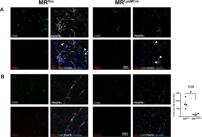
Fig. 5.
Immunofluorescence micrographs of heart sections from old MRflox and MRLysMCre mice showing A macrophages (CD68+ cells), fibroblasts (PDGFRα+ cells), and TIMD4 immunoreactivity. Arrows indicate CD68+ and TIMD4+ cells. In the aged heart of MRflox mice, TIMD4– macrophages were localized predominantly to areas containing a large amount of fibroblasts. B Immunoreactivity for CCL2 in CD68 and PDGFRα-positive areas. Old hearts from MRLysMCre mice displayed a weak expression of CCL2 in PDGFRα-positive areas. Nuclei were stained with NucBlue™. Mean ± SEM, n = 4 per group; *p < 0.05

