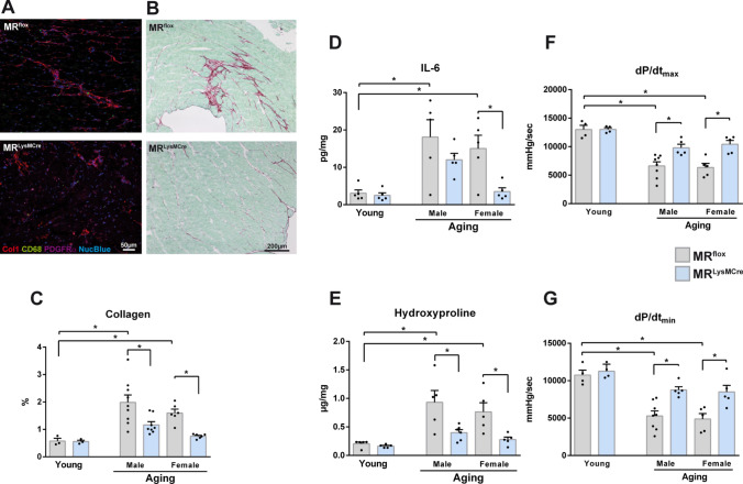
Fig. 6.
MR deficiency in macrophages protects mice from cardiac inflammation, fibrosis, and heart dysfunction induced by aging. A Immunofluorescence micrographs of heart sections from old MRflox and MRLysMCre mice showing macrophages (CD68+ cells), fibroblasts (PDGFRα+ cells), and collagen type 1 immunoreactivity. B Sirius red/fast green staining of representative heart sections from old MRflox and MRLysMCre mice. C Interstitial fibrosis quantified by picrosirius red polarization microscopy, D cardiac IL-6 protein and E hydroxyproline levels in young and old MRflox and MRLysMCre mice. F Left ventricular maximal rate of pressure rise (dP/dtmax) and G maximal rate of pressure decline (dP/dtmin) measured in vivo with a conductance catheter in young and old MRflox and MRLysMCre mice. Aged MRflox and MRLysMCre mice were on average 22 (± 0.7) months old. Mean ± SEM, n = 3–9 per group; *p < 0.05

