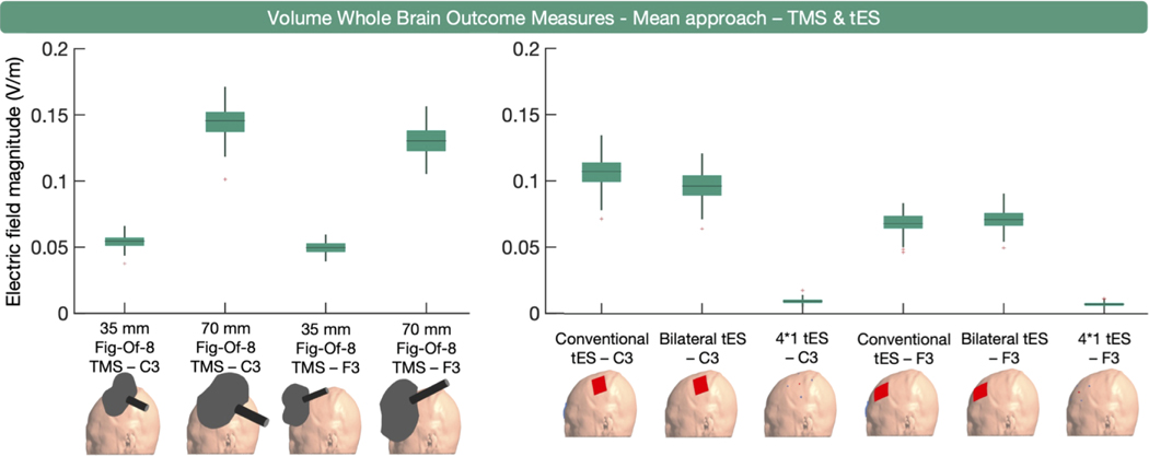
Fig. 3.
Volume-level, whole brain, mean E-field magnitude outcome measure per modality. On the left, the boxplots for TMS indicate that the larger 70 mm figure- eight TMS coil induces the highest E-field magnitude. On the right, the boxplots for tES indicate that conventional and bilateral tES induce substantially higher mean E-field magnitudes than 4 × 1 tES. Overall, the mean E-field magnitudes are higher when targeting C3 compared to F3.

