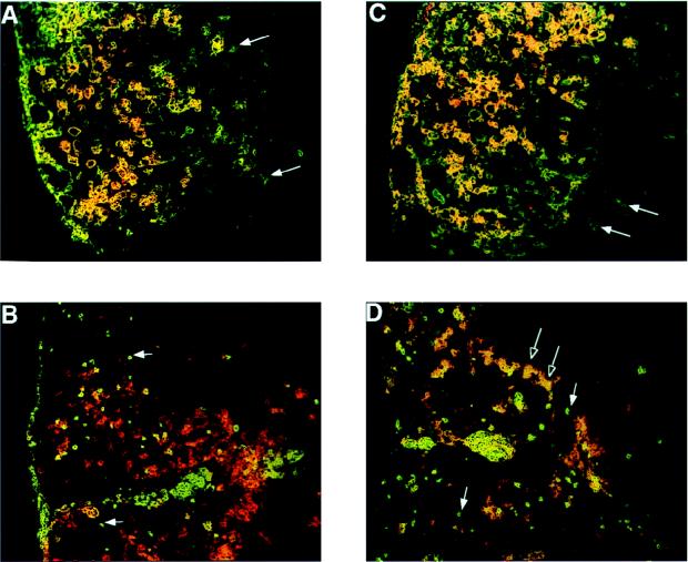
FIG. 3.
Demonstration of B7-1 and B7-2 in the spinal cords of TMEV-infected mice by immunohistochemistry. Frozen sections (5 to 6 μm) of SJL/J spinal cord were prepared 45 days p.i. and stained sequentially for F4/80 or CD4 (green), followed by B7-1 or B7-2 (red). B7-1 (A) and B7-2 (C) colocalize on large F4/80+ macrophages and microglia with the exception of a few F4/80+ microglia at the edges of the lesions (arrows). B7-1 and B7-2 do not colocalize to small CD4+ T cells in the central part of the lesions (B and D, filled arrows), but a subpopulation of large, blast-like CD4+ T cells at the lesion margins express B7-2 (D, open arrows).

