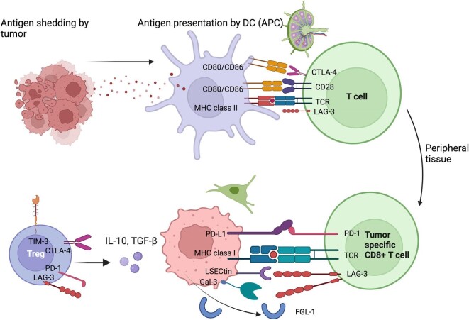
Figure 1:
Tumor-associated antigens are presented by APC in secondary lymphoid organs to T cells. Activation of T cells is tightly regulated by immune checkpoint of which CTLA-4 in the most important proximally. In peripheral tissues and at tumoral level PD-1/ PD-L1 pathways exert an inhibitory role on T cells and promotes cancer survival. Tumoral cells also express liver sinusoidal endothelial cell lectin (LSECtin), Galectin-3 (Gal-3), and fibrogen-like protein-1 (FGL-1) that bind LAG-3 to induce T cell anergy. Tumor survival and growth is further enhanced by the presence of tumor-infiltrating regulatory T cells that are known to express higher levels of CTLA-4, PD-1, LAG-3, and T cell immunoglobulin and mucin domain-containing protein 3 (TIM-3) and to secrete higher levels of IL-10 and TGF-beta, promoting tumoral tolerance within the tumoral microenvironment.

