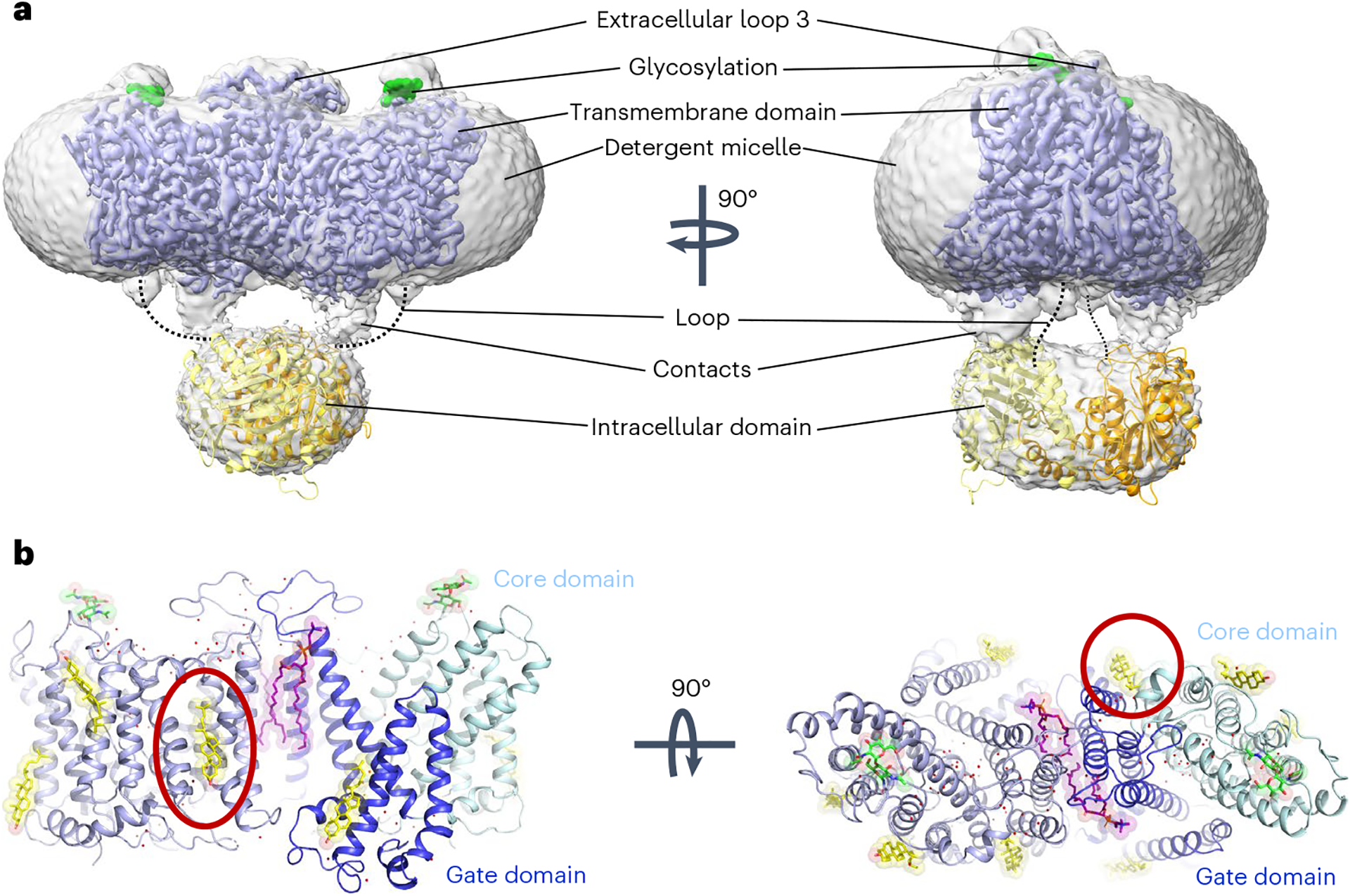
Fig. 1 |. Cryo-EM structure of full-length human AE1/SLC4A1.

a, Cryo-EM density of overall AE1 homodimer and detergent micelle (gray), overlaid with density of the membrane domain of AE1 (mdAE1, light blue). Glycosylation sites are highlighted in green, and the crystal structure of the cytoplasmic domain (cdAE1, PDB 1HYN) homodimer (yellow/orange) is loosely fit into the density. Dotted lines highlight that the loop connecting cdAE1 and mdAE1 termini is in a different position from noncovalent contacts observed between the domains. b, mdAE1 structure (light blue) including bound phospholipids (purple) and cholesterol (yellow), glycosylation sites (green) and water molecules (red spheres). Gate and core domains of one of the protomers have been highlighted in dark blue and pale cyan, respectively. Presumed inhibitory cholesterol located between domains is encircled in red.
