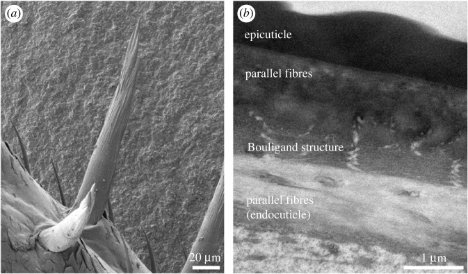
Figure 4.
Chitin–protein fibre organization in the sensory spines of the terrestrial isopod Porcellio. (a) A scanning electron micrograph of sensory spines on the walking leg. (b) A transmission electron micrograph of the spine cuticle in longitudinal section. While the fibres in the outer exocuticle are parallel, those in the inner exocuticle form the Bouligand structure. In the endocuticle, fibres are again parallel.

