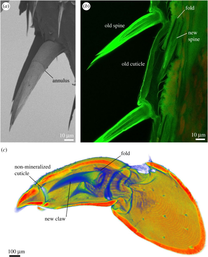
Figure 7.
The formation of skeletal elements affects their structure. (a) A scanning electron micrograph of a sensory spine on an isopod leg with a clearly visible annulus. The image was obtained as described in Vittori et al. [35]. (b) Formation of the spines during preparation for moult in an optical section obtained with confocal microscopy. As new sensory spines are constructed, they are folded in an epidermal pocket. As they extend after moult, the position of the fold remains visible as the annulus. The leg was processed and imaged as described in Vittori et al. [65] using a Leica Stellaris 8 confocal microscope. (c) Formation of claws on the legs in the isopod Ligia visualized using micro-CT. The image was obtained as described in Vittori et al. [40] using a NeoScan N80 micro-CT device. Just like sensory spines, the claw forms in an invagination that remains visible as a ring of non-mineralized cuticle when the claw is extended. Is this simply the result of the claw's formation or does it perform a mechanical function?

