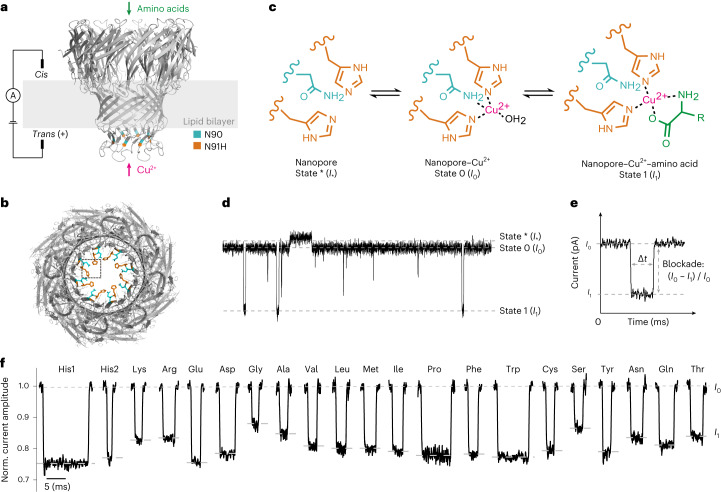
Fig. 1. Experimental setup and principle of amino acid detection.
a, Schematic of the experimental setup. Amino acids and copper ions were added to the cis and trans chambers, respectively. A voltage of +50 mV was applied during measurement. The N91H substitutions in eight subunits are highlighted in orange. b, Bottom-view structure of the MspA-N91H nanopore (predicted using SWISS-MODEL). The dotted box shows a binding site for the copper ion. c, Proposed sensing mechanism. Two adjacent histidine residues (position 91) and one asparagine residue (position 90) coordinate a copper ion. Then, the α-amine and α-carboxyl groups of the amino acid coordinate the copper–histidine complex. d, A representative current trace showing the corresponding current change for three binding states. e, Illustration of current blockade induced by the binding of amino acid. f, Representative signals of current blockade events of 20 amino acids. The events of histidine exhibited two populations, His1 and His2.

