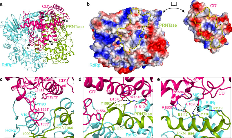
Fig. 6. L–L interaction.
a Overview of the CD domain (CD′) of the second L binding to the RdRp and PRNTase domains of the intact L. Three arrows represent the views shown in (c–e). b The surfaces of CD′ and RdRp-PRNTase are colored by electrostatic potential from red to blue (negative to positive, respectively). The interface of each is indicated by a dashed circle. c–e Magnified view of the interactions between CD′ and RdRp-PRNTase. The polar interactions are indicated by dashed lines.

