Figure 2.
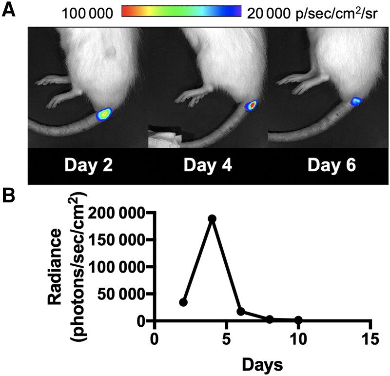
Optical imaging of the rat vertebral discitis-osteomyelitis model using bioluminescent Staphylococcus aureus. A, S aureus Xen 29 was inoculated at the third intervertebral space followed by serial imaging on a Xenogen IVIS 50 instrument as described in the Materials and Methods. Shown are representative bioluminescent signals at the site of inoculation on days 2, 4, and 6, indicative of technically successful intervertebral inoculation. B, Quantitative analysis of bioluminescent imaging performed at 2, 4, 6, 8, and 10 days demonstrates that day 4 is the optimal time for bioluminescent imaging of positron emission tomography tracers.

