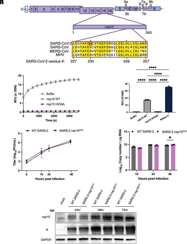
Fig. 1.
Construction of recombinant SARS-CoV-2 with inactive nsp15mut endoribonuclease. (A) Diagram of the SARS-CoV-2 genome. Full-length nsp15 of SARS-CoV-2 is shown in the center. Sequence alignment among betacoronaviruses, from Top to Bottom: SARS-CoV-2 USA-WA1/2020, SARS-CoV, MERS-CoV EMC/2012, and MHV strain A59. Conserved residues are shown in yellow with the catalytic histidine residues of SARS-CoV-2, H234, and H249, designated with asterisks. Nsp15 mutation site for SARS-CoV-2 nsp15mut generated in this study, H234, shown in red. (B) SARS-CoV-2 WT, nsp15mut, and buffer alone catalytic activity measured by dequenching of 6-FAM upon ssRNA cleavage measured in relative fluorescence units recorded at 5-min intervals over a 60-min period. (C) Total quantified ssRNA cleavage measured for WT nsp15, nsp15mut, buffer alone, and RNase A at the end of 1 h. (D–F) VeroE6 cells were infected with either WT or nsp15mut SARS-CoV-2 (MOI 0.1 in D and MOI 1 in E and F). (D) Supernatants were collected at indicated times post infection and titered via plaque assay. Data shown are the average of two independent experiments. (E) Intracellular RNA was collected at the indicated time points post infection and genome copy number per μg RNA was quantified by RT-qPCR using a standard curve and primers directed against SARS-CoV-2 RdRp (nsp12). (F) Cells were lysed at indicated time points using protein lysis buffer. Samples were separated via SDS-PAGE and transferred to a PVDF membrane for immune detection with antibodies against SARS-CoV-2 nsp15, SARS-CoV-2 N, and GAPDH.

