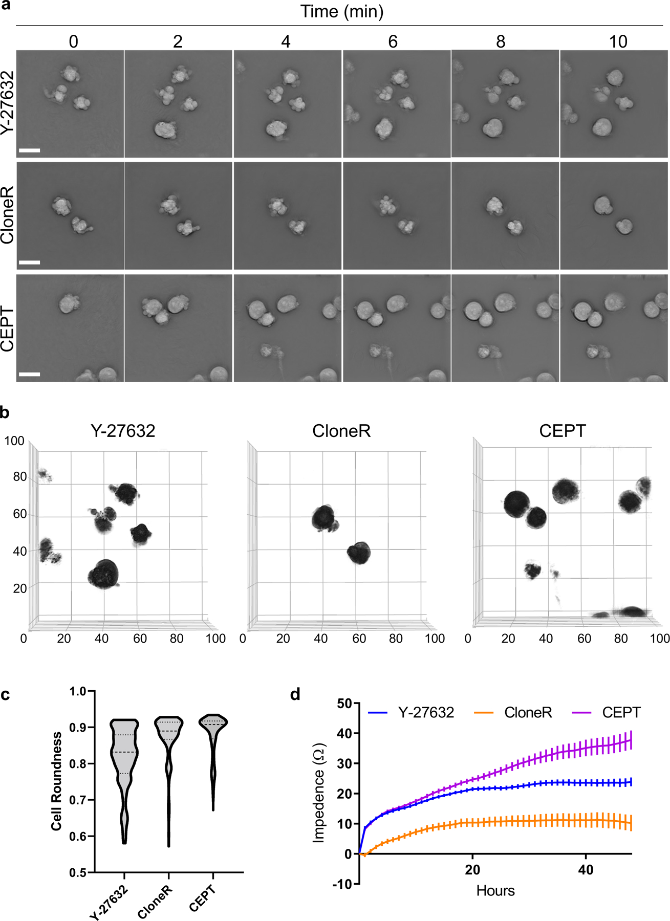
Fig. 2: Cell morphology and cell adhesion profile of dissociated hPSCs after treatment with Y-27632, CloneR, or CEPT.

a, Label-free live-cell imaging (Nanolive) showing the first 10 min after plating dissociated cells (WA09). Abnormal phenotypes are caused by cell contractions and membrane blebbing in the presence of Y-27632 (10 µM) and CloneR (1X), whereas treatment with fast-acting CEPT mitigates cell stress and ensures circularity of cell bodies. b, Analysis of membrane blebbing and cell roundness of single cells at 10 min after cell dissociation and treatment with Y-27632 (10 µM), CloneR (1X), or CEPT. Images represent 3D renditions of 60–70 z-planes stacked together. A customized fully automated digital image analysis algorithm segmented each cell as an individual object and measured the shape. Cell roundness is quantified per cell as deviation from perfect mathematical roundness. Six fields of view were analyzed for each condition. Note that CloneR has less cells (black circles) in the field of view because the addition of CloneR (1:10 dilution as recommended by the manufacturer) changes the density of the medium and the cells do not settle to the bottom of the plate as fast as they do with Y-27632 and CEPT. c, Violin plots of live-cell images (3D Cell Explorer) showing distribution of cell roundness and membrane blebbing at 10 min post-treatment with Y-27632, CloneR, or CEPT. Data are from n ≥ 230 for each condition. d, Cell adhesion time-course measured by impedance analysis of cells passaged with Y-27632 (10 µM), CloneR (1X), or CEPT. Data are mean ± s.e.m.; n = 16 wells for each group. Scale bars, 20 µm (a and b); x and y-axes in µm.
