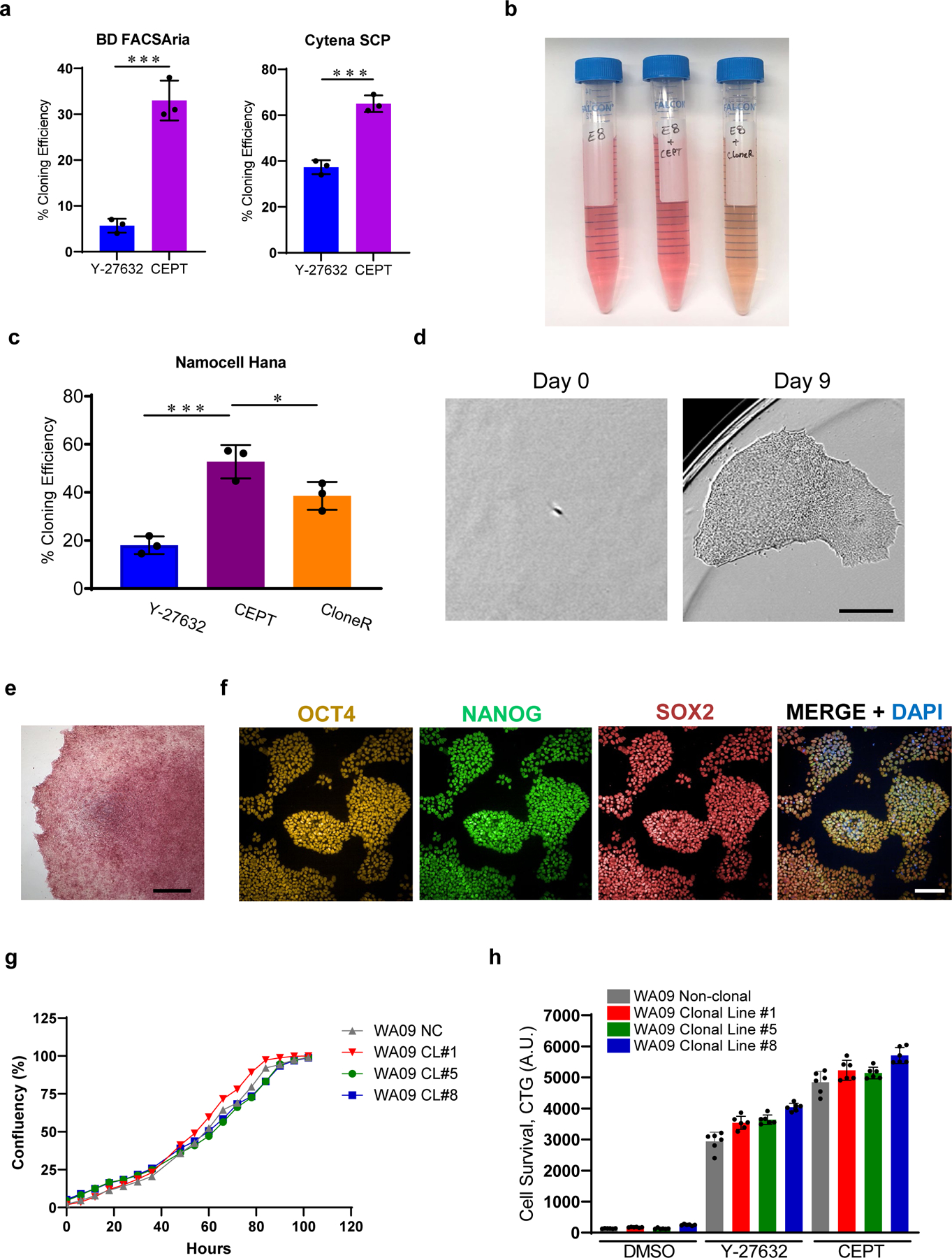
Fig. 3: CEPT improves single-cell cloning efficiency and maintains pluripotency.

a, Single-cell cloning efficiency in the presence of Y-27632 (10 µM) and CEPT as tested on two different platforms including single-cell printing with Cytena (Molecular Devices), and cell sorting with FACSAria (Becton Dickinson). Figure adapted with permission from ref. 21. b, E8 Medium after supplementation with CEPT or CloneR. Note that addition of CloneR (1:10 dilution according to manufacturer) changes the color of cell culture medium. c, Quantification of single-cell cloning efficiency (WA09) using microfluidics-based cell sorting (Namocell Hana) after treatment with Y-27632 (10 µM), CloneR (1X), or CEPT. d, Phase-contrast images showing a single cell (WA09) that survived dissociation and generated a clonal colony in 9 days. e, Representative image of a clonal colony (WA09) expressing pluripotency-associated marker alkaline phosphatase. f, Representative immunocytochemical images showing that clonal cell lines maintain pluripotency and express OCT4, NANOG and SOX2 (clonal cell line #1 from WA09). g, Image-based analysis comparing cell growth rate of non-clonal parental line (NC) and derived clonal cell lines 1, 5, and 8 (CL #1, 5 and 8). h, Luminescent cell viability assay showing that the sensitivity to enzymatic cell dissociation is comparable in non-clonal parental line and derived clonal cell lines 1, 5, and 8. Scale bars, 300 µm (d), 400 µm (e), and 100 µm (f). Data are mean ± s.d.; n = 3 96-well plates for each group (a, c), *P ≤0.05, **P ≤0.01, ***P ≤0.001, two-way ANOVA and Student’s t-test, respectively. Data are mean ± s.d.; n ≥ 3 (g, h).
