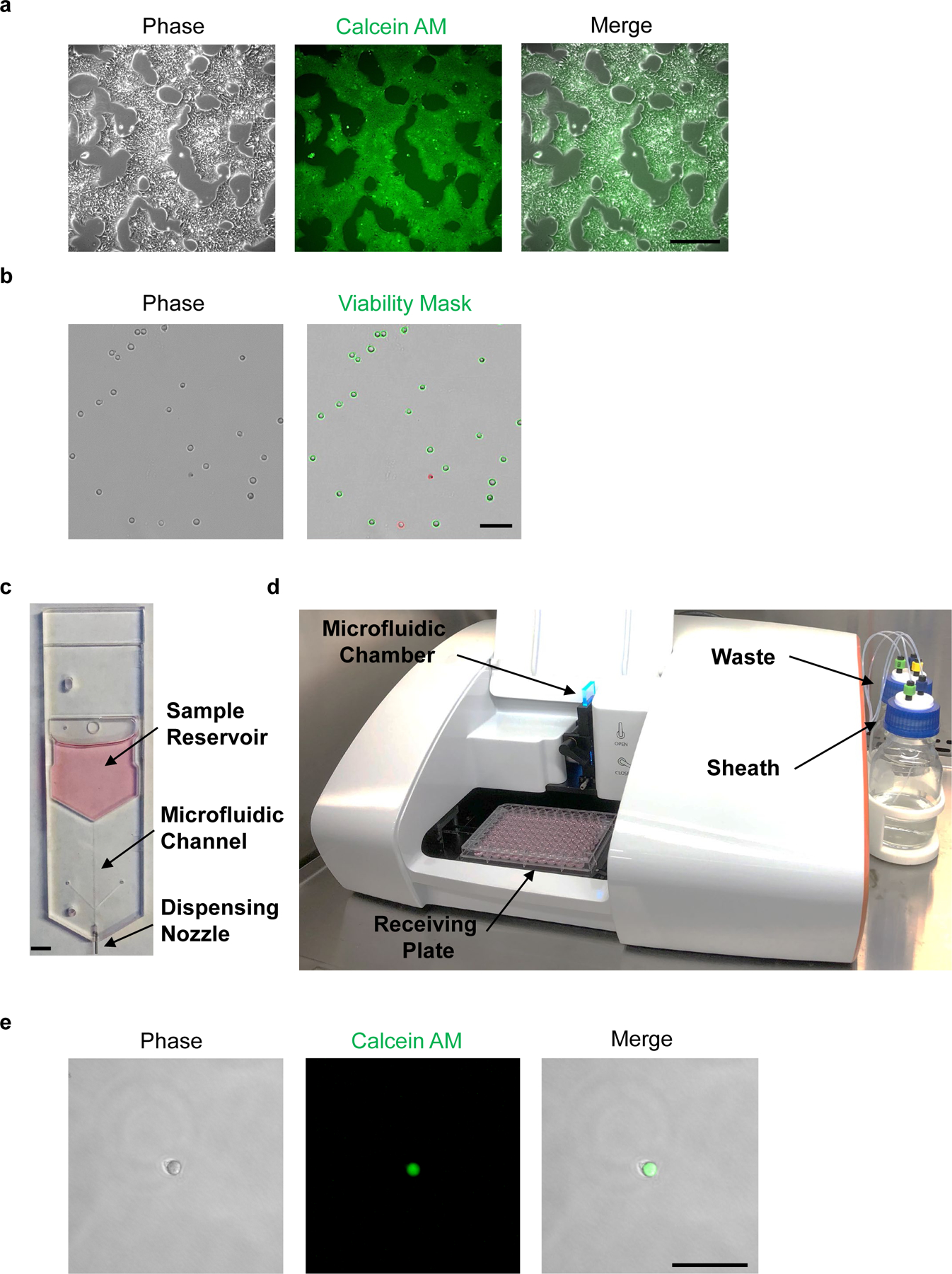
Fig. 5. Live-cell staining and microfluidic single-cell dispensing of hPSCs.

a, Phase-contrast image of hPSCs before Calcein AM staining (left panel). Fluorescence microscopic analysis of hPSCs at 1 h post-staining with Calcein AM (center and right panels). b, Cell suspension of dissociated hPSCs should avoid clumps and yield low frequency of doublets (left panel). Trypan blue exclusion test can be used to measure cell viability; live and dead cells are shown with green or red outline, respectively (right panel). c, Disposable microfluidic cell cartridge depicting sample reservoir, microfluidic channel, and dispensing nozzle. d, Hana (Namocell) single-cell dispenser with microfluidic chamber, receiving plate, waste and sheath bottles. e, Fluorescence microscopic image of a single calcein-labeled hPSC after microfluidic dispensing in a 1 μl droplet. Scale bars, 400 µm (a), 200 µm (b and e) and 5 mm (c).
