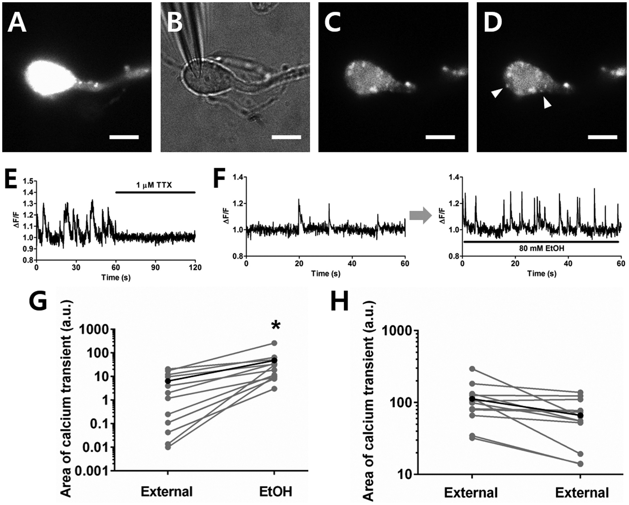
Fig. 7. Calcium imaging shows that calcium entry is increased in presynaptic boutons after EtOH incubation.

A. Fluorescence image after calcium dye loading in a mechanically-dissociated neuron. B. Wide-field image of patch pipette with giga-ohm seal on the neuron. C. Fluorescence image 2 min after opening the membrane for whole-cell recording. D. Calcium transients were observed from a subpopulation of dye-loaded puncta (white arrowheads) after diluting the dye in the postsynaptic neuron. Scale bar = 10 μm. E. The spontaneous calcium transients were suppressed by TTX. F. Representative spontaneous normalized Ca2+ transients before and during EtOH incubation. G. The spontaneous calcium entry increased in the presence of 80 mM EtOH (paired t-tests *P < 0.05, n = 12). H. In control experiments without EtOH exposure, no increase of calcium entry was observed. The small decrease in transients in the control condition is likely due to photo bleaching (n = 12).
