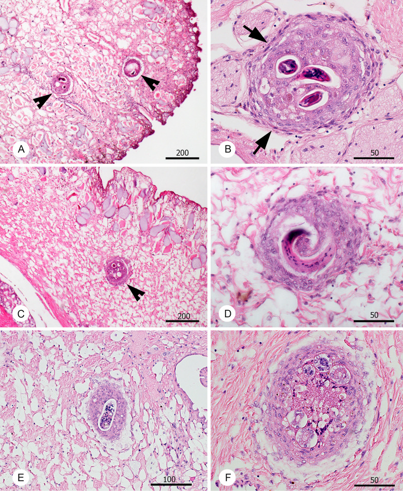
Fig. 1.
Histopathological changes related to the presence of L3 larvae of A. cantonensis (arrowheads) in the tissues of experimental slugs infected in experiment A2 and examined 45 dpi. Veronicella cubensis (A–B) and V. sloanei (C–F). The nodules surrounding the L3 larvae in case of V. cubensis are surrounded by a layer of fibroblasts (arrows) and filled with mixed inflammatory cells (B). The nodules surrounding A. cantonensis L3 larvae in V. sloanei were less compact with minimum fibroblasts (D). Notably, numerous larvae were found dead in V. sloanei, partly (E) or almost entirely disintegrated (F). All scale bars in μm.

