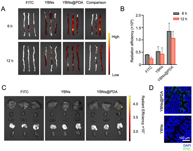
Fig. 3.
YBNs@PDA localizes in inflamed colon in DSS-treated mice. (A, B) After 6 h and 12 h of treating animals with 10 mg/kg free-FITC, YBNs-FITC or YBNs@PDA-FITC, colons were imaged by in vivo imaging system (IVIS) (A) and quantified for FITC fluorescence signal (B). Comparison picture was taken by comparing one colon sample from each group. (C) Ex vivo fluorescence images of major organs collected from colitis mice 12 h after oral gavage of free-FITC, YBNs-FITC or YBNs@PDA-FITC, respectively. (D) CLSM images sections of the distal colon tissues from mice orally administered with YBNs-FITC or YBNs@PDA-FITC

