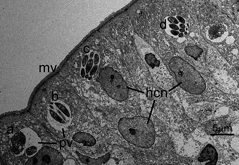
Fig. 1.
Transmission electron micrograph of C. cayetanensis tissue stages in a duodenal biopsy. Tissue stages (a–d) are located within cytoplasmic parasitophorous vacuoles (pv) at the apical end of infected enterocytes. Note the intact overlying microvillous border (mv) and host cell nuclei (hcn). Four schizonts (a–d) are visible in this section; the schizont in (a) is immature, while the schizonts in (b–d) are mature and contain fully formed merozoites. Note the small size (~5 μm) of the schizonts.

