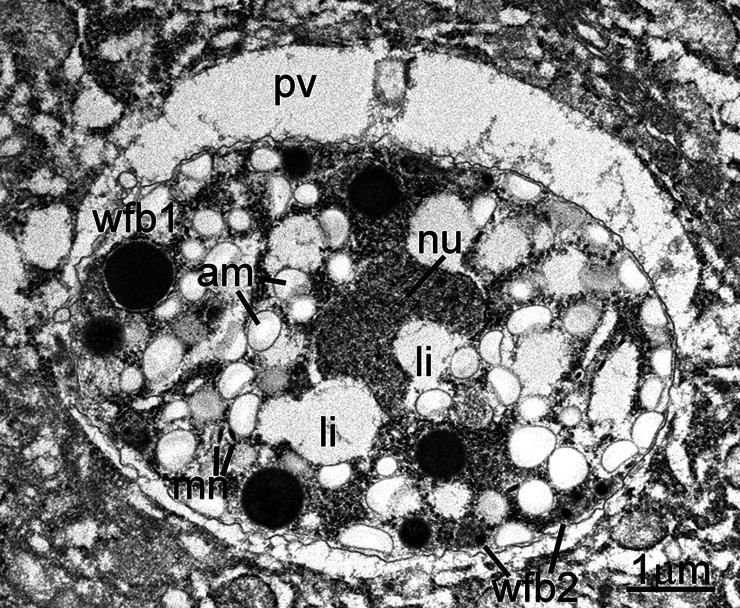
Fig. 10.
Transmission electron micrograph of a C. cayetanensis macrogamont (female) within a parasitophorous vacuole (pv) in a duodenal biopsy. This 6.8 × 4.8 μm macrogamont is at a more advanced stage than the macrogamont shown in Fig. 9. Note an elongated nucleus (nu), numerous amylopectin granules (am), few lipid bodies (li) and peripheral wall-forming bodies (wfb1 and wfb2).

